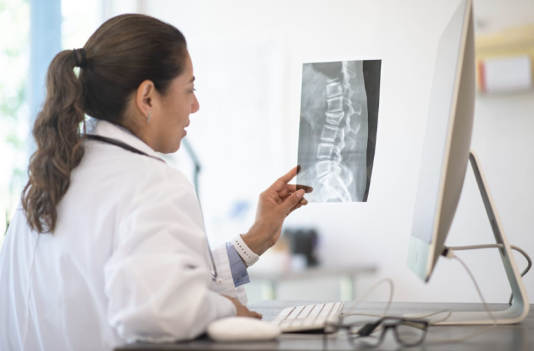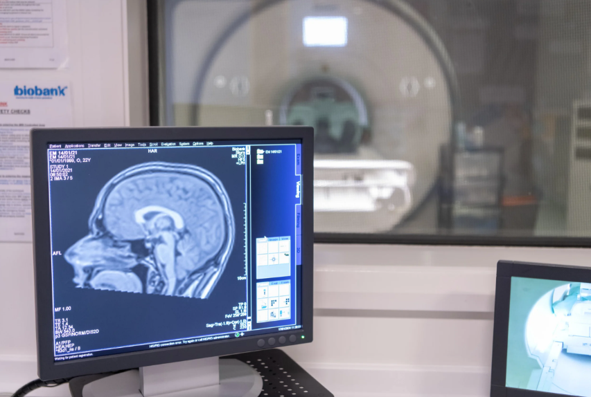

Neuroradiology plays a significant role in detecting and diagnosing brain tumors through advanced imaging techniques. Exams such as magnetic resonance imaging (MRI) help healthcare professionals identify abnormal growths, assess their size and location, and evaluate their effects on surrounding brain structures. Here is more information on how neuroradiology detects and diagnoses brain tumors:
Detecting Brain Tumors With Imaging
MRI and CT scans are the two primary imaging tools in neuroradiology. Each method produces detailed images of the brain, but each uses different technology to create them. Your doctor chooses a scan based on your symptoms, medical history, and the information needed.
How Do MRI Scans Work?
MRI uses a strong magnetic field and radio waves to create soft-tissue images. It does not use radiation, and it produces highly detailed views of brain structures. A technologist may inject a contrast dye, a substance that makes certain tissues stand out more clearly, during the procedure.
See also: Understanding the Benefits of Choosing an Out-of-Network Spine Specialist
How Do CT Scans Work?
CT combines a series of X-rays taken from different angles to build cross-sectional images. It works faster than MRI, so emergency departments often use it as a first step. Although CT shows less soft-tissue detail, it clearly shows bleeding, swelling, and bone changes. Some patients receive CT when metal implants or other factors prevent an MRI.
Assessing Tumor Size and Characteristics
When imaging reveals an abnormal area, the neuroradiologist studies it in detail. This review produces a written report, and your physician uses it to discuss the findings with you. The report often includes the following key features:
- Size: Measurements in millimeters or centimeters show how large the growth is and allow comparison over time.
- Location: The specific brain region helps explain certain symptoms, such as changes in speech or movement.
- Borders: Smooth or irregular edges show how the growth relates to nearby tissue.
- Contrast enhancement: How an area absorbs contrast dye provides information about its blood supply.
- Surrounding effects: Swelling or pressure on nearby structures shows how the growth affects the brain.
Differentiating Tumors From Other Conditions
Not every abnormal area on a scan is a tumor. Infections, strokes, multiple sclerosis lesions, and blood vessel abnormalities sometimes look similar on imaging. Neuroradiology specialists use their training to distinguish among these conditions, reducing diagnostic uncertainty.
Advanced MRI techniques support this process. Diffusion-weighted imaging measures how water moves through tissue, and perfusion imaging shows blood flow to a specific area. MR spectroscopy, which analyzes tissue chemistry, adds another layer of detail. These tools provide a more comprehensive picture than a standard scan. Your medical history also shapes interpretation. Recent illness, prior surgery, or a known condition gives context, so the neuroradiologist reviews this information alongside the images.
Guiding Testing and Treatment Planning
Neuroradiology findings often shape the next steps in your care. If a scan suggests a tumor, your doctor may recommend a biopsy, a procedure that removes a small tissue sample for laboratory testing. Neuroradiology helps identify the safest and most accessible location for that sample.
Surgeons, oncologists, and radiation specialists also rely on detailed images. Surgeons use them to map a path that avoids key brain areas, and radiation teams use them to define treatment zones. In some cases, cerebral angiography, an imaging test of the brain’s blood vessels, provides extra information before surgery.
Schedule Your Neuroradiology Appointment Today
Neuroradiology plays a fundamental role in diagnosing, planning, and monitoring treatment for neurological conditions. Advanced imaging techniques like MRI and CT provide detailed insights to guide precision care. Follow-up imaging makes sure treatments are tracked and adjusted as needed for the best outcomes. Contact a modern neuroradiology imaging practice to schedule your appointment today.